Download
1 / 25
290 likes | 783 Views
Heart Sounds . Dr. Maha Alenazy. Objectives . Understand types of heart sounds How to examine heart for sounds Understand murmurs Use phonocarciography. What are heart sounds?.

E N D
Heart Sounds Dr. MahaAlenazy
Objectives Understand types of heart sounds How to examine heart for sounds Understand murmurs Use phonocarciography
What are heart sounds? • The heart sounds are the noises (sound) generated by the beating heart and the resultant flow of blood through it • In cardiac auscultation, an examiner uses a stethoscope to listen for these sounds, which provide important information about the condition of the heart.
What are the different heart sounds? • Lub (first heart sound (S1)) which is associated with the closure of the AV valves • Dub (second heart sound (S2)) which is associated with the closure of the semilunar valves
Slapping of the valves leaflets is not enough to generate a heart sound. • The causes of the 1st heart sound: • During systole the AV valves are closed & blood tries to flow back to the atrium back bulging the AV valves. But the taut chordae tendinae stop the back bulging and causes the blood to flow forward. • This in addition to the actual closure of the valves will cause vibration of the valves, blood & the walls of the ventricles which is presented as the 1st heart sound
The causes of the 2nd heart sound: • During diastole, blood in the blood vessels tried to flow back to the ventricles cause the semilunar valves to bulge. But the elastic recoil of the arteries cause the blood to bounce forward which will vibrate the blood the valves and the ventricle walls. • This also in addition to the actual closure of the semilunar valves are presented as the 2nd heart sound.
More heart sounds • The 3rd heart sound: is the heard in the mid diastole due to the blood that fills the ventricles. • The 4th heart sound: also known as atrial heart sound. It occurs when the atrium contracts & pumps blood to the ventricles. This sound is almost never heard by the stethoscope.
Where to listen? • Tricuspid valve: is best heard at the Rt half the lower end of the sternum body • Mitral valve: is best heard at the Apex of the heart (Lt 5th intercostal space at the mid-clavicular line). Here S1 is louder than S2. • Pulmonary valves: is best heard at the Lt medial 2ndintercostalspace (in this position, S2 is louder than S1) • Aortic valve: is best heard in the medial 2ndRtinetercostal space.
Physiological splitting of S2 • 1. Chest wall expands during inspiration • 2. Intrathoracic pressure becomes more negative to form a vacuum • 3. Venous return from the body to the right heart increases + Venous return from the lungs to the left heart decreases • 4. Right ventricular volume and emptying time increases + Left ventricular volume and emptying time decreases • 5. Pulmonic valve closure is delayed + Aortic valve closure is advanced • 6. S2 splits into A2 and P2respectively • 7. Expiration equalizes filling and emptying times on both sides of the heart eliminating the splitting of S2
Splitting of S1 • Very narrow not usually heard. Mitral and tricuspid valves have 20-30msec difference. • Best heard at the lower left sternal border with the stethoscope diaphragm (T1 but M1 at apex) EXPERTS.
Abnormal splitting S1: heard • The split may be increased to 60 msecin: • A. Electrical problems: • patients with right bundle branch block, Ebstein's anomaly, or other conditions in which there is electrical delay in activation of one of the two ventricles (e.g., ventricular ectopic beats, ventricular tachycardia, AV block with idioventricular rhythm, and left ventricular pacing).
Cont. • B. Mechanical delays: • As in in closure of the mitral valve (e.g., mitral stenosis and left atrial myxoma) or the tricuspid valve (e.g., right atrial myxoma) may cause abnormal splitting of the first heart sound
Where to listen for the Split? A split S1 heart sound is best heard at the tricuspid listening post since T1 is much softer than M1. (Rthalf the lower end of the sternum body) A split S2 is best heard at the pulmonic valve listening post sine P2 is much softer than A2. (the Lt medial 2ndIS)
Murmurs • generated by turbulent flow of blood, which may occur inside or outside the heart. Murmurs may be physiological (benign) or pathological (abnormal). • Different murmurs are audible in different parts of the cardiac cycle, depending on the cause of the murmur.
THRILL • Is a vibration felt by the examiner on palpitation. • Happens in severe heart problems.
Types of Murmur Systolic Diastolic Continous
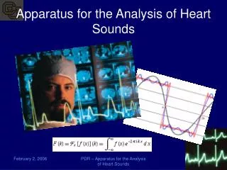
Phonocardiography • diagnostic technique that creates a graphic record, or phonocardiogram, of the sounds and murmurs produced by the contracting heart, including its valves and associated great vessels.
The phonocardiogram is obtained either with a chest microphone or with a miniature sensor in the tip of a small tubular instrument that is introduced via the blood vessels into one of the heart chambers.
The phonocardiogram usually supplements the information obtained by listening to body sounds with a stethoscope (auscultation) and is of special diagnostic value when performed simultaneously with measurement of the electrical properties of the heart (electrocardiography) and pulse rate.
http://library.med.utah.edu/kw/pharm/hyper_heart1.html http://www.cvphysiology.com/Heart%20Disease/HD002.htm http://www.learntheheart.com/PDF2-heartsounds.pdf