Download
1 / 33
330 likes | 555 Views
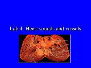
Lab 4: Heart sounds and vessels. Overview. Review Heart sounds Blood supply to the heart Blood vessels Veins Portal systems New lab website: http://isu.indstate.edu/~jowhitak/cmritzi.htm Practical lab review: Sunday 2-5 pm. Review.

E N D
Overview • Review • Heart sounds • Blood supply to the heart • Blood vessels • Veins • Portal systems • New lab website: http://isu.indstate.edu/~jowhitak/cmritzi.htm • Practical lab review: Sunday 2-5 pm
Review • How do volume and pressure in the ventricles change during atrial contraction? • What region of the heart controls the electrical signal through the heart? • Which standard limb lead has a negative lead on the right arm and a positive lead on the left arm?
Overview • Review • Heart sounds • Blood supply to the heart • Blood vessels • Veins • Portal systems
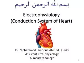
Heart sounds“lubb-dubb” • S1: longer and louder • S2: softer and shorter • S3: found in children and juveniles • S1 & S2 occur in conjunction with the opening and closing of valves • Actually caused by turbulence of blood
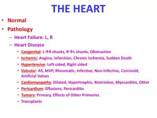
Valvular troubles • Valvular Insufficiency: valve failure causing backwards flow • Any defect in the valves can lead to heart failure • Defective valves can be replaced with artificial valves or with pig valves
Valvular Troubles (cont) • Mitral valve prolapse: tri- or bicuspid cusps bulge back into the atrium. • Symptoms: chest pain, fatique, shortness of breath • Cause: hereditary • Valvular Stenosis: cusps of the valve are stiff and opening is constricted by scar tissue • Results from autoimmune disease • Causes enlarged heart • Blood moving backwards through the valves causes a heart murmur
Blood supply to the heart muscles • The endocardium prevents blood from seeping through the heart • How is blood supplied to the myocardium and the pericardium? • Coronary arteries originating from the aortic arch
Arteries in the heart Aorta Right Coronary Left Coronary Marginal artery Circumflex Artery Posterior IV Artery Anterior IV Artery Anastomoses
Point where two arteries join to reach a common destination If one artery becomes blocked, the other can take blood to the appropriate destination Anastomoses
Veins in the heart Greater Cardiac Vein Small Cardiac Vein Middle Cardiac Vein Coronary Sinus Right Atrium
Contributes to over 710,000 heart attack and stroke, and peripheral vascular disease deaths each year. Fatty blockage in coronary artery Artherosclerosis
Artherosclerosis • Cause: Abundance of low-density lipids and defective receptors for LDL in the coronary arteries. • LDL: cholesterol, free fatty acids and phospholipids • Arterial cells with defective receptors will take in too much cholesterol • Results in obstruction of the arterial lumen
Cardiac Ischemia and Infarction • Ischemia: Loss of blood flow • Infarction: Death of myocardial cells; heart attack • Cause: Artheriosclerosis blocks a cardiac artery. The downstream region does not receive enough oxygen causing cell death. The death of these cells weakens the heart wall disrupting electrical pathways leading to fibrillation.
Overview • Review • Heart sounds • Blood supply to the heart • Blood vessels • Veins • Portal systems
Blood Vessels Arteries Capillaries Veins
Tunica externa: loose connective tissue Tunica media: smooth muscle, elastin, collagen Tunica interna: endothelium Structure of Blood Vessels
Types of Arteries • Conducting (elastic) • Passively accommodate blood flow • Distributing (muscular) • High smooth muscle content • Deliver blood to specific organs • Arterioles • Control blood flow • Link to capillaries
Pre-capillary sphincters Open and close capillary beds ¾ of all capillaries are ususally closed Structure: endothelium only Capillaries
Continuous: uninterrupted tube Only allow small solutes such as glucose to enter Blood-brain barrier Fenestrated: riddled with pores Allow rapid passage of small molecules Hold proteins in the vessels Important in organs that engage in rapid filtration Types of Capillaries
Veins • Venules: Collect blood from capillaries • Porous • Veins: • Lower pressure than the arteries • Thinner walls (little tunica media or externa) but larger lumens • Walls expand easy and accomadate more blood than arteries
How does blood travel in veins? 1. Messaging action of skeletal muscle 2. Pressure gradient in the body: • Positive pressure in the abdominal cavity and negative in the thoracic cavity draws blood up
3.Venous valves prevent backflow Failure of valves results in varicose veins Venous Valves
Special Circulations: Portal Systems Artery • Portal systems: Blood flows between two consecutive capillary beds before returning to the heart Capillary 1 Capillary 2 Vein
Connects capillaries of the intestines to capillaries of the liver Delivers newly absorbed nutrients to the liver Hepatic Portal System
Connects hypothalamus to pituitary gland Responsible for transporting hormones Hypothalamo-hypophyseal portal system
Overview • Review • Heart sounds • Blood supply to the heart • Blood vessels • Valves • Portal systems • Cat heart and human heart
Cat Dissection • Finish identifying arteries and veins • Remove cat heart and dissect in half • Valves and chambers • Chordae Tendinai • Coronary Arteries