Download
1 / 31
330 likes | 411 Views
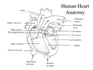
Heart Anatomy. Approximately the size of your fist Wt. = 250-300 grams Location In the mediastinum between the lungs Superior surface of diaphragm ⅔’s of it lies to the left of the midsternal line Anterior to the vertebral column, posterior to the sternum. Heart Anatomy. Figure 18.1.

E N D
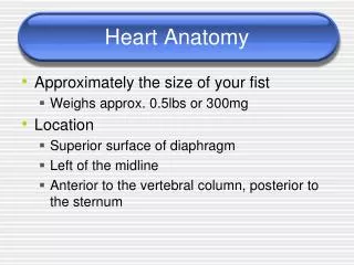
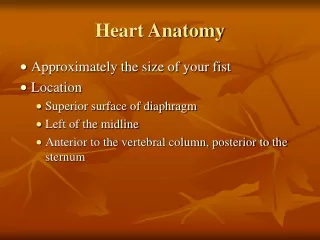
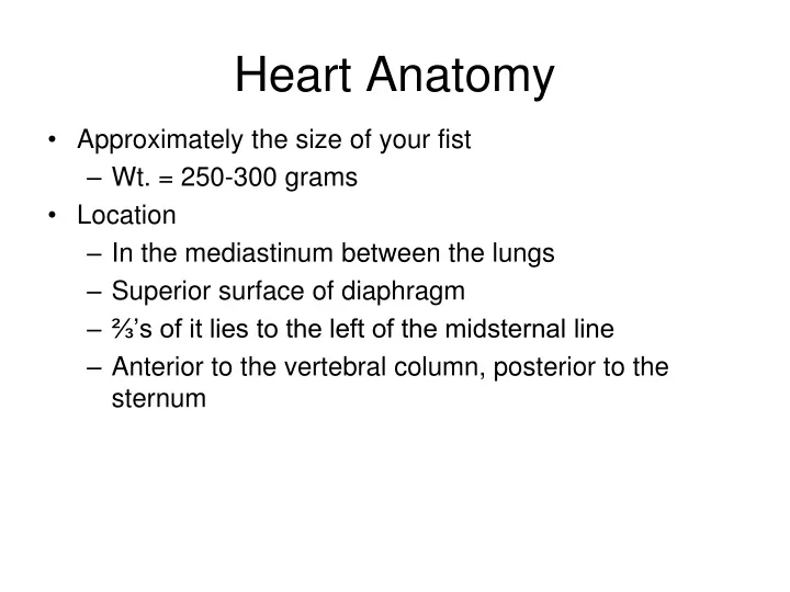
Heart Anatomy • Approximately the size of your fist • Wt. = 250-300 grams • Location • In the mediastinum between the lungs • Superior surface of diaphragm • ⅔’s of it lies to the left of the midsternal line • Anterior to the vertebral column, posterior to the sternum
Heart Anatomy Figure 18.1
Coverings of the Heart • Pericardium – a double-walled sac around the heart • Composed of: • A superficial fibrous pericardium • A deep two-layer serous pericardium • The parietal layer lines the internal surface of the fibrous pericardium • The visceral layer or epicardium lines the surface of the heart • They are separated by the fluid-filled pericardial cavity. • Protects and anchors the heart • Prevents overfilling of the heart with blood • Allows for the heart to work in a relatively friction-free environment
Pericardial Layers of the Heart Figure 18.2
Layers of the Heart Wall • Epicardium – visceral pericardium • Myocardium – cardiac muscle layer forming the bulk of the heart • Endocardium – endothelial layer of the inner myocardial surface
Heart Anatomy • External markings • Apex - pointed inferior region • Base - upper region • Coronary sulcus • Indentation that separates atria from ventricles • Anterior and posterior interventricular sulcus • Separates right and left ventricles • Internal divisions • Atria (superior) and ventricles (inferior) • Interventricular and interatrial septa
Atria of the Heart • Atria - receiving chambers of the heart • Receive venous blood returning to heart • Separated by an interatrial septum (wall) • Foramen ovale - opening in interatrial septum in fetus • Fossa ovalis - remnant of foramen ovale • Each atrium has a protruding auricle • Pectinate muscles mark atrial walls • Pump blood into ventricles • Blood enters right atria from superior and inferior venae cavae and coronary sinus • Blood enters left atria from pulmonary veins
Gross Anatomy of Heart: Frontal Section Figure 18.4e
Ventricles of the Heart • Ventricles are the discharging chambers of the heart • Papillary muscles and trabeculae carneae muscles mark ventricular walls • Separated by an interventricular septum • Contains components of the conduction system • Right ventricle pumps blood into the pulmonary trunk • Left ventricle pumps blood into the aorta • Thicker myocardium due to greater work load • Pulmonary circulation supplied by right ventricle is a much low pressure system requiring less energy output by ventricle • Systemic circulation supplied by left ventricle is a higher pressure system and thus requires more forceful contractions
External Heart: Anterior View Figure 18.4b
Structure of Heart Wall • Left ventricle – three times thicker than right • Exerts more pumping force • Flattens right ventricle into a crescent shape Figure 18.7
Heart Valves • Heart valves ensure unidirectional blood flow through the heart • Composed of an endocardium with a connective tissue core • Two major types • Atrioventricular valves • Semilunar valves • Atrioventricular (AV) valves lie between the atria and the ventricles • R-AV valve = tricuspid valve • L-AV valve = bicuspid or mitral valve • AV valves prevent backflow of blood into the atria when ventricles contract • Chordae tendineae anchor AV valves to papillary muscles of ventricle wall • Prevent prolapse of valve back into atrium
Semilunar Heart Valves • Semilunar valves prevent backflow of blood into the ventricles • Have no chordae tendinae attachments • Aortic semilunar valve lies between the left ventricle and the aorta • Pulmonary semilunar valve lies between the right ventricle and pulmonary trunk • Heart sounds (“lub-dup”) due to valves closing • “Lub” - closing of atrioventricular valves • “Dub”- closing of semilunar valves
Fibrous Skeleton • Surrounds all four valves • Composed of dense connective tissue • Functions • Anchors valve cusps • Prevents overdilation of valve openings • Main point of insertion for cardiac muscle • Blocks direct spread of electrical impulses
Conducting System • Cardiac muscle tissue has intrinsic ability to: • Generate and conduct impulses • Signal these cells to contract rhythmically • Conducting system • A series of specialized cardiac muscle cells • Sinoatrial (SA) node sets the inherent rate of contraction
Innervation • Heart rate is altered by external controls • Nerves to the heart include: • Visceral sensory fibers • Parasympathetic branches of the vagus nerve • Sympathetic fibers – from cervical and upper thoracic chain ganglia
External Heart: Posterior View Figure 18.4d
Major Vessels of the Heart • Vessels returning blood to the heart include: • Superior and inferior venae cavae • Open into the right atrium • Return deoxygenated blood from body cells • Coronary sinus • Opens into the right atrium • Returns deoxygenated blood from heart muscle (coronary veins) • Right and left pulmonary veins • Open into the left atrium • Return oxygenated blood from lungs
Major Vessels of the Heart • Vessels conveying blood away from the heart include: • Pulmonary trunk • Carries deoxygenated blood from right ventricle to lungs • Splits into right and left pulmonary arteries • Ascending aorta • Carries oxygenated blood away from left atrium to body organs • Three major branches • Brachiocephalic • Left common carotid, • Left subclavian artery
Blood Flow Through the Heart Figure 18.6
Pathway of Blood Through the Heart and Lungs Figure 18.5
Coronary Circulation • Coronary circulation • The functional blood supply to the heart muscle itself • R and L Coronary arteries are 1st branches off the ascending aorta • Coronary sinus (vein) empties into R. atrium • Collateral routes ensure blood delivery to heart even if major vessels are occluded
Coronary Circulation - Arteries • Right Coronary Artery • Supplies blood to • Right atrium and posterior surface of both ventricles • Branches into the • Marginal artery - extends across surface of R. ventricle • Posterior interventricular artery • Found in posterior interventricular sulcus • Left Coronary Artery • Supplies blood to • Left atrium and left ventricle • Branches into • Circumflex artery • Anterior interventricular artery • Found in anterior interventricular sulcus • Connected with posterior interventricular artery via arterial anastomoses
Coronary Circulation: Arterial Supply Figure 18.7a
Coronary Circulation - Veins • Coronary sinus - • Vein that empties into right atrium • Receives deoxygenated blood from: • Great cardiac vein - on anterior surface • Posterior cardiac vein • Drains area served by circumflex • Middle cardiac vein • Drains area served by posterior interventricular artery • Small cardiac vein • Drains blood from posterior surfaces of right atrium and ventricle
Coronary Circulation: Venous Supply Figure 18.7b
Microscopic Anatomy of Heart Muscle • Cardiac muscle cells • Short, striated, branched, and interconnected • The connective tissue endomysium acts as both tendon and insertion • Intercalated discs anchor cardiac cells together and allow free passage of ions • Heart muscle behaves as a functional syncytium • Many mitochondria (25% of total volume)
Microscopic Anatomy of Heart Muscle Figure 18.11
Disorders of the Heart • Coronary artery disease • Atherosclerosis – fatty deposits • Arteriosclerosis - hardening of the arteries • Angina pectoris – chest pain • Myocardial infarction – blocked coronary artery • Silent ischemia – no pain or warning • Fibrillation - irregular heart beat; may occur in either atria or ventricles