Download
1 / 30
401 likes | 738 Views
HEART ANATOMY. The Heart Wall. serous membrane. A serous membrane consists of a single layer of flattened mesothelial cells applied to the surface of a thin layer of collagenous tissue that attaches to underlying fascia.

E N D
serous membrane • A serous membrane consists of a single layer of flattened mesothelial cells applied to the surface of a thin layer of collagenous tissue that attaches to underlying fascia. • The mesothelium of the serous membrane forms the lining of a closed serous membrane cavity. • Serous membrane lining the wall of a serous cavity is designated parietal while that covering viscera is called visceral. • Connecting serous membrane runs between parietal and visceral components.
The myocardium. Note the endocardial layer, which consists of endothelium supported by a rather thick layer of subendocardial connective tissue (green). The ventricular lumen is indicated. Valve Lumen
HEART ANATOMY (EXTERNAL VIEW) • The heart is a complex muscular pump that maintains oxygen and blood circulation through the lungs and the rest of the body through Both the systemic and the pulmonary cerculations. • The heart pumps about 7200 liters/day. • Is about the size of you clenched fist
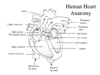
Internal Anatomy and Organization • Atria – separated by the interatrial septum • Ventricles – separated by the interventricular septum • Atrioventricular valves extend into the openings between the atria and ventricles permitting the blood to flow in one direction only. • Contraction of the papillary muscles prevent the atrioventricular valves from folding back into the atria.
Right Atrium • Receives blood from the superior vena cava (head, neck upper limbs, chest) inferior vena cava (rest of the trunk, lower limbs, and viscera), and coronary veins of the heart through the coronary sinus. • Oval impression – Fossa Ovalis • Contains pectinate muscles with trabecule
The distinction between the posterior Smooth walled and the anterior trabeculated appendage. The junction between the two is marked by The formed muscle bundle, the crista terminalis. The trabeculae tend to run at right angles to the crista. The inside of the right atrial chamber presents a The posterior surface, a septal surface, and an anterior surface. The floor of the chamber can be considered as tricuspid valve orifice orientated obliquely to the right The inferior vena cava opens into the junction of the posterior wall And the floor. The Right Atrium Ant CT Post FO Septal IVC
Right Atrium, Extensive trabecular pouch found beneath the orifice of the inferior vena cava(the so-called sub-eustachian sinus). IVC SES http://www.rjmatthewsmd.com/Definitions/anatomy_ofthe_heart.htm
Right Ventricle • Blood flows from the right atrium to the right ventricle through the cusps of the right atrioventricular valve known as the tricuspid valve. • The tricuspid valve is attached by long tendons called chordae tendineae to the papillary muscles. • When the right ventricle contracts, the tricuspid closes preventing blood from entering the right atrium. The chordae tendineae keep the tricuspid from folding back into the right atrium. • Blood exits the right ventricle through the conus anteriosus as the pulmonary semilunar valve opens into the pulmonary trunk dividing into the right and left pulmonary arteries leading to the lungs.
PT AVS CT PM The Right ventricle MB
Left Atrium • Smaller than right atrium • Thicker walls than right atrium • 2 left & 2 right pulmonary veins • Oval impression – Fossa Ovalis • Atrial Appendage (longer & narrower) • Receives oxygen rich blood from the two right and two left pulmonary veins. • Blood passes from the left atrium to the left ventricle through the left atrioventricular valve or bicuspid.
Left Atrium • The left atrium is rather smaller than the right, but its walls are thicker, measuring about 3 mm it consists, like the right, • of two parts, a principal cavity and an auricula. • The principal cavity is cuboidal in form, and concealed, in front, by the pulmonary artery and aorta; in front and to the right it is separated from the right atrium by the atrial septum; opening into it on either side are the two pulmonary veins. • The auricula is somewhat constricted at its junction with the principal cavity; it is longer, narrower, and more curved than that of the right side, and its margins are more deeply indented. It is directed forward and toward the right and overlaps the root of the pulmonary artery. 35
Left Ventricle • Oval shaped • Larger than right • Walls 3 X thicker than right • Smooth walls • Papillary muscles • Cordae tendinae • Contractions causes the bicuspid to close keeping the blood from backing up in the left atrium; distance between the apex and base increases; diameter of the ventricle chambers decrease. • Blood exits through the semilunar valve into the ascending aorta. • Right and left coronary arteries originate at the aortic sinuses and deliver blood to the heart. • Blood passes into the descending aorta and into the systemic circuit.
Valves of the HeartThe heart has four valves for: Pumping action of the heart. Maintaining unidirectional blood flow.
PT AVS CT PM Tricuspid Valve
2 triangular leaflets Larger, thicker, stronger than tricuspid Anterior leaflet (aortic or septal) Posterior leaflet (ventricular) Papillary muscle – contraction occurs during systole to shorten Cordae Tendinae prevent MR during ventricular systole 3 triangular shaped leaflets Names Anterior Septal Posterior Papillary muscles & chordae tendinae are present but play a more important role in the high pressure chamber of LV Mitral Valve Tricuspid valve
Pulmonary semi-lunar valve Lt. Atrium Aortic semi-lunar valve
3 Semi-lunar cusps Semi lunar shape Attached to a fibrous ring within the wall of pulmonary trunk Orientation: Left anterior Right anterior Posterior 3 Similar to pulmonary Leaflets - 3 Semi lunar shape Attached to a fibrous ring within the wall of wall of aortic artery Orientation Anterior Left posterior Right posterior Aortic valve Pulmonary valve
Diastole Systole