Download

1 / 52
580 likes | 912 Views
Heart Anatomy. Approximately the size of a fist Location In the mediastinum between second rib and fifth intercostal space On the superior surface of diaphragm Two-thirds to the left of the midsternal line Enclosed in pericardium, a double-walled sac. PLAY. Animation: Rotatable heart.

E N D
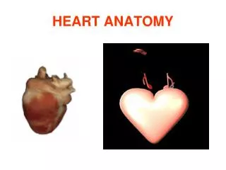
Heart Anatomy • Approximately the size of a fist • Location • In the mediastinum between second rib and fifth intercostal space • On the superior surface of diaphragm • Two-thirds to the left of the midsternal line • Enclosed in pericardium, a double-walled sac PLAY Animation: Rotatable heart
Midsternal line 2nd rib Sternum Diaphragm Point of maximal intensity (PMI) (a) Figure 18.1a
Aorta Superior vena cava Parietal pleura (cut) Pulmonary trunk Left lung Pericardium (cut) Apex of heart Diaphragm (c) Figure 18.1c
Pericardium • Superficial fibrous pericardium • Protects, anchors, and prevents overfilling
Pericardium • Deep two-layered serous pericardium • Separated by fluid-filled pericardial cavity (decreases friction)
Pulmonary trunk Fibrous pericardium Parietal layer of serous pericardium Pericardium Pericardial cavity Myocardium Epicardium (visceral layer of serous pericardium) Heart wall Myocardium Endocardium Heart chamber Figure 18.2
Layers of the Heart Wall • Epicardium—visceral layer of the serous pericardium
Layers of the Heart Wall • Myocardium • Spiral bundles of cardiac muscle cells • Fibrous skeleton of the heart: crisscrossing, interlacing layer of connective tissue • Anchors cardiac muscle fibers • Supports great vessels and valves • Limits spread of action potentials to specific paths
Layers of the Heart Wall • Endocardium is continuous with endothelial lining of blood vessels
Pulmonary trunk Fibrous pericardium Parietal layer of serous pericardium Pericardium Pericardial cavity Myocardium Epicardium (visceral layer of serous pericardium) Heart wall Myocardium Endocardium Heart chamber Figure 18.2
Cardiac muscle bundles Figure 18.3
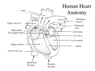
Chambers • Four chambers • Two atria • Separated internally by the interatrial septum • Coronary sulcus (atrioventricular groove) encircles the junction of the atria and ventricles • Auricles increase atrial volume
Chambers • Two ventricles • Separated by the interventricular septum • Anterior and posterior interventricular sulci mark the position of the septum externally
Left common carotid artery Brachiocephalic trunk Left subclavian artery Superior vena cava Aortic arch Ligamentum arteriosum Right pulmonary artery Left pulmonary artery Ascending aorta Left pulmonary veins Pulmonary trunk Right pulmonary veins Auricle of left atrium Circumflex artery Right atrium Left coronary artery (in coronary sulcus) Right coronary artery (in coronary sulcus) Anterior cardiac vein Left ventricle Right ventricle Great cardiac vein Right marginal artery Anterior interventricular artery (in anterior interventricular sulcus) Small cardiac vein Inferior vena cava Apex (b) Anterior view Figure 18.4b
Atria: The Receiving Chamberson diagram • Vessels entering right atrium • Superior vena cava • Inferior vena cava • Coronary sinus • Vessels entering left atrium • Right and left pulmonary veins
Ventricles: The Discharging Chambers on diagram • Walls are ridged by trabeculae carneae • Papillary muscles project into the ventricular cavities • Vessel leaving the right ventricle • Pulmonary trunk • Vessel leaving the left ventricle • Aorta
Aorta Left pulmonary artery Superior vena cava Left atrium Right pulmonary artery Left pulmonary veins Pulmonary trunk Right atrium Mitral (bicuspid) valve Right pulmonary veins Fossa ovalis Aortic valve Pectinate muscles Pulmonary valve Tricuspid valve Left ventricle Papillary muscle Right ventricle Interventricular septum Chordae tendineae Trabeculae carneae Epicardium Inferior vena cava Myocardium Endocardium (e) Frontal section Figure 18.4e
Pathway of Blood Through the Heart • The heart is two side-by-side pumps • Right side is the pump for the pulmonary circuit • Vessels that carry blood to and from the lungs • Left side is the pump for the systemic circuit • Vessels that carry the blood to and from all body tissues
Capillary beds of lungs where gas exchange occurs Pulmonary Circuit Pulmonary veins Pulmonary arteries Aorta and branches Venae cavae Left atrium Left ventricle Right atrium Heart Right ventricle Systemic Circuit Oxygen-rich, CO2-poor blood Capillary beds of all body tissues where gas exchange occurs Oxygen-poor, CO2-rich blood Figure 18.5
Pathway of Blood Through the Heart • Right atrium tricuspid valve right ventricle • Right ventricle pulmonary semilunar valve pulmonary trunk pulmonary arteries lungs PLAY Animation: Rotatable heart (sectioned)
Pathway of Blood Through the Heart • Lungs pulmonary veins left atrium • Left atrium bicuspid valve left ventricle • Left ventricle aortic semilunar valve aorta • Aorta systemic circulation PLAY Animation: Rotatable heart (sectioned)
Pathway of Blood Through the Heart • Equal volumes of blood are pumped to the pulmonary and systemic circuits • Pulmonary circuit is a short, low-pressure circulation • Systemic circuit blood encounters much resistance in the long pathways • Anatomy of the ventricles reflects these differences
Left ventricle Right ventricle Interventricular septum Figure 18.6
Coronary Circulation • The functional blood supply to the heart muscle itself • Arterial supply varies considerably and contains many anastomoses (junctions) among branches • Collateral routes provide additional routes for blood delivery
Coronary Circulation don’t copy • Arteries • Right and left coronary (in atrioventricular groove), marginal, circumflex, and anterior interventricular arteries • Veins • Small cardiac, anterior cardiac, and great cardiac veins
Aorta Superior vena cava Left pulmonary artery Right pulmonary artery Right pulmonary veins Left pulmonary veins Auricle of left atrium Right atrium Left atrium Inferior vena cava Great cardiac vein Coronary sinus Right coronary artery (in coronary sulcus) Posterior vein of left ventricle Posterior interventricular artery (in posterior interventricular sulcus) Left ventricle Apex Middle cardiac vein Right ventricle (d) Posterior surface view Figure 18.4d
Homeostatic Imbalances • Angina pectoris • Thoracic pain caused by a fleeting deficiency in blood delivery to the myocardium • Cells are weakened • Myocardial infarction (heart attack) • Prolonged coronary blockage • Areas of cell death are repaired with noncontractile scar tissue
Heart Valves • Ensure unidirectional blood flow through the heart • Atrioventricular (AV) valves • Prevent backflow into the atria when ventricles contract • Tricuspid valve (right) • Mitral valve (left) • Chordae tendineae anchor AV valve cusps to papillary muscles
Heart Valves • Semilunar (SL) valves • Prevent backflow into the ventricles when ventricles relax • Aortic semilunar valve • Pulmonary semilunar valve
Pulmonary valve Myocardium Aortic valve Tricuspid (right atrioventricular) valve Area of cutaway Mitral valve Tricuspid valve Mitral (left atrioventricular) valve Myocardium Tricuspid (right atrioventricular) valve Aortic valve Mitral (left atrioventricular) valve Pulmonary valve Aortic valve Pulmonary valve Aortic valve Pulmonary valve Area of cutaway (b) Fibrous skeleton Mitral valve Tricuspid valve (a) Anterior Figure 18.8a
Myocardium Tricuspid (right atrioventricular) valve Mitral (left atrioventricular) valve Aortic valve Pulmonary valve Pulmonary valve Aortic valve Area of cutaway (b) Mitral valve Tricuspid valve Figure 18.8b
Pulmonary valve Aortic valve Area of cutaway Mitral valve Tricuspid valve Chordae tendineae attached to tricuspid valve flap Papillary muscle (c) Figure 18.8c
Opening of inferior vena cava Mitral valve Chordae tendineae Tricuspid valve Myocardium of right ventricle Myocardium of left ventricle Pulmonary valve Aortic valve Area of cutaway Papillary muscles Mitral valve Interventricular septum Tricuspid valve (d) Figure 18.8d
1 Blood returning to the heart fills atria, putting pressure against atrioventricular valves; atrioventricular valves are forced open. Direction of blood flow Atrium Cusp of atrioventricular valve (open) 2 As ventricles fill, atrioventricular valve flaps hang limply into ventricles. Chordae tendineae 3 Atria contract, forcing additional blood into ventricles. Papillary muscle Ventricle (a) AV valves open; atrial pressure greater than ventricular pressure Atrium 1 Ventricles contract, forcing blood against atrioventricular valve cusps. Cusps of atrioventricular valve (closed) 2 Atrioventricular valves close. Blood in ventricle 3 Papillary muscles contract and chordae tendineae tighten, preventing valve flaps from everting into atria. (b) AV valves closed; atrial pressure less than ventricular pressure Figure 18.9
Aorta Pulmonary trunk As ventricles contract and intraventricular pressure rises, blood is pushed up against semilunar valves, forcing them open. (a) Semilunar valves open As ventricles relax and intraventricular pressure falls, blood flows back from arteries, filling the cusps of semilunar valves and forcing them to close. (b) Semilunar valves closed Figure 18.10
Heart Sounds • Two sounds (lub-dup) associated with closing of heart valves • First sound occurs as AV valves close and signifies beginning of systole • Second sound occurs when SL valves close at the beginning of ventricular diastole • Heart murmurs: abnormal heart sounds most often indicative of valve problems usually the valve not closing all the way.
Aortic valvesounds heard in 2nd intercostal space at right sternal margin Pulmonary valve sounds heard in 2nd intercostal space at left sternal margin Mitral valvesounds heard over heart apex (in 5th intercostal space) in line with middle of clavicle Tricuspid valvesounds typically heard in right sternal margin of 5th intercostal space Figure 18.19
Mechanical Events: The Cardiac Cycle • Cardiac cycle: all events associated with blood flow through the heart during one complete heartbeat • Systole—contraction • Diastole—relaxation
Other Factors that Influence Heart Rate • Age • Gender • Exercise • Body temperature
Developmental Aspects of the Heart don’t copy • Embryonic heart chambers • Sinus venous • Atrium • Ventricle • Bulbus cordis
Ductus arteriosus Aorta Arterial end Arterial end Superior vena cava 4a Pulmonary trunk 4 Tubular heart Ventricle Foramen ovale 3 Atrium 2 Ventricle 1 Ventricle Inferior vena cava Venous end Venous end (a) Day 20: Endothelial tubes begin to fuse. (b) Day 22: Heart starts pumping. (c) Day 24: Heart continues to elongate and starts to bend. (d) Day 28: Bending continues as ventricle moves caudally and atrium moves cranially. (e) Day 35: Bending is complete. Figure 18.23
Developmental Aspects of the Heart • Fetal heart structures that bypass pulmonary circulation • Foramen ovale connects the two atria • Ductus arteriosus connects the pulmonary trunk and the aorta
Developmental Aspects of the Heart • Congenital heart defects • Lead to mixing of systemic and pulmonary blood • Involve narrowed valves or vessels that increase the workload on the heart
Narrowed aorta Occurs in about 1 in every 500 births Occurs in about 1 in every 2000 births Occurs in about 1 in every 1500 births (c) Tetralogy of Fallot. Multiple defects (tetra = four): (1) Pulmonary trunk too narrow and pulmonary valve stenosed, resulting in (2) hypertrophied right ventricle; (3) ventricular septal defect; (4) aorta opens from both ventricles. (a) Ventricular septal defect. The superior part of the inter- ventricular septum fails to form; thus, blood mixes between the two ventricles. More blood is shunted from left to right because the left ventricle is stronger. (b) Coarctation of the aorta. A part of the aorta is narrowed, increasing the workload of the left ventricle. Figure 18.24