Download
1 / 1
10 likes | 171 Views
Neu5Ac2-O-3Gal1-O-4GlcNAc1-O-2Man1. O. 6. Man11-O-4GlcNAc1-O-4GlcNAc-ol. Neu5Ac2-O-3Gal1-O-4GlcNAc1. 3. O. 4 2. O. Man1. O. Neu5Ac2-O-6Gal1-O-4GlcNAc1. Applications of Glycosidase in Glycoproteins Structure Determination by AP-MALDI MS/MS Analysis

E N D
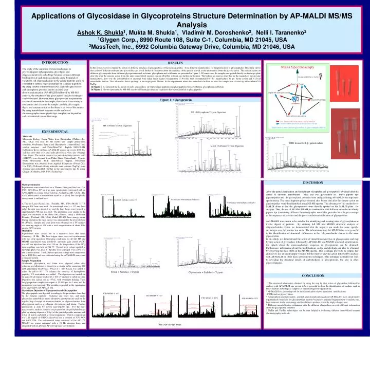
Neu5Ac2-O-3Gal1-O-4GlcNAc1-O-2Man1 O 6 Man11-O-4GlcNAc1-O-4GlcNAc-ol Neu5Ac2-O-3Gal1-O-4GlcNAc1 3 O 4 2 O Man1 O Neu5Ac2-O-6Gal1-O-4GlcNAc1 Applications of Glycosidase in Glycoproteins Structure Determination by AP-MALDI MS/MS Analysis Ashok K. Shukla1, Mukta M. Shukla1, Vladimir M. Doroshenko2, Nelli I. Taranenko2 1Glygen Corp., 8990 Route 108, Suite C-1, Columbia, MD 21045, USA 2MassTech, Inc., 6992 Columbia Gateway Drive, Columbia, MD 21046, USA INTRODUCTION RESULTS In this poster, we have studied the action of different enzymes on glycoproteins or their glycopeptides from different manufactures for the purification of glycopeptides. This study shows that action of different endo and exo glycosidase can reveal further in formation about the sequence of the protein as well as the information about the glycosylation. The enzyme action on different glycopeptides from different glycoproteins such as fetuin, glycophorin and ovalbumin are presented in figure 1. IN some cases the samples are spotted directly on the target plate after the after the enzyme action from the mini immobilized enzyme column (TopTip) without any further purification. The buffers are used as described in the manuals of the enzyme manufacturer, how ever the concentration of enzymes were taken much higher concentration (5-10 fold) than recommended by the manufacturer to get faster action and to avoid nonvolatile buffers. This allowed to direct spotting at the target plate. Elution. In the experiments where the nonvolatile buffers are used the sample was cleaned up with carbon+C18 NuTip. In Figure 1. we demonstrate the action of endo- glycosidase on tryptic digest peptides and glycopeptides from ovalbumin, glycophorin and fetuin. In Figure 2. shows representative MS-MS data for different glycopeptide fragments that were identified as glycopeptides. The study of the sequence of monosaccharides in glycoconjugates (glycoproteins, glycolipids and oligosaccharides) is a challenge because so many different binding sites at each monosaccharide cause thousands of variations. All oligosaccharides in the acidic fractions could be converted to neutral oligosaccharides by sialidase digestion. By using soluble or immobilized exo- and endo-glycosidase and atmospheric pressure matrix-assisted laser desorption/ionization (AP-MALDI) followed by MS-MS analysis, the structure of the glyco part of the glycoconjugate can be obtained. However, these glycoproteins are present in very small amounts in the sample, therefore it is necessary to concentrate and clean-up the sample carefully after tryptic digest and enzyme action so that there is no loss of the sample. By using immobilized enzymes on the surface of chromatographic micro pipette tips, samples can be purified and concentrated in nanoliter range. TopTip Figure 1: Glycoprotein NuTip MS of Glycophorin Trypsin Digestion MS of Fetuin Trypsin Digestion EXPERIMENTAL Materials Molecular Biology Grade Water from Biowittaker (Walkersville, MD, USA) was used for the matrix and sample preparation solutions. Ovalbumin, Fetuin and Glycophorin , immobilized and soluble enzymes and ProteoMassTM Peptide MALDI-MS Calibration Kit to calibrate AP-MALDI spectra up to m/z 4000 Da, sialidase and other exo- and endo-glycosidase were also obtained from Sigma. The matrix material, α-cyano-4-hydroxycinnamic acid (4-HCCA) was obtained from Fluka (Buch, Switzerland). Trypsin beads (Poroszyme Bulk Immobilized Trypsin, PerSeptive Biosystems) was obtained from Applied Biosystems (Foster City, CA, USA). Different affinity materials mini columns (TopTip) were obtained and embedded (NuTip) in the micropipette tips by using Glygen ( Columbia, MD, USA) Technology. . OTD treated with b-galactosidase MS of Ovalbumin Trypsin Digestion DISCUSSION Mass spectrometry Experiments were carried out on a Thermo Finnigan (San Jose, CA, USA) LCQ Deca XP ion trap mass spectrometer integrated with an AP/MALDI ion source (MassTech Inc., Columbia, MD, USA). The AP/MALDI source is described in detail in ref. [8-9], but our specific arrangement is outlined here. A Thermo Laser Science Inc. (Franklin, MA, USA) Model 337 Si nitrogen UV laser was used. Its wavelength was l = 337 nm, laser pulse duration was about 4 ns, and the laser beam was focused to approximately 500 mm size spot. The maximum laser energy on the target was measured to be about 140 mJ/pulse, using a Molectron Detector (Portland, OR, USA) Model EM-400 laser energy meter. During operation, the laser energy was attenuated to the level of about 60 mJ/pulse. Sample and laser spots were observed on a TV monitor at a viewing angle of 450 with a total magnification of about 100x using a CCD camera. Operation Experiments were carried out in a repetitive laser shot mode (frequency 10 Hz). The laser trigger times were not synchronized with the LCQ operation. Operating conditions for all full MS and MS/MS experiments were as follows: automatic gain control (AGC) was off, ion injection time was 220 ms, the temperature of the LCQ input capillary was held at 280 0C. Typical high voltage applied to the target plate was 2000 V. Spectra were averaged over a 30 s to 3 min collection time. The LCQ was operated in high mass range mode (up to 4000 Da), and was calibrated using the AP/MALDI source and a standard protein. Sample Preparation Ovalbumin, glycophorin and fetuin were digested either after reduction and alkylation or directly in a volatile buffer containing 100 mM ammonium bicarbonate. 3.0 µl of 1 mM CaCl2 was added to adjust the pH to 8.5. To enhance the recovery of hydrophobic peptides, 5 % acetonitrile was added. The digestion was carried out by using 10 µl trypsin beads with 1:100 v/v enzyme to substrate ratio. Digestion was carried out at +37oC, with overnight shaking. Then, the glycoprotein samples were spun at 15,000 rpm for 3 min. and the supernatant was removed. The peptides generated in the supernatant were analyzed by AP-MALDI MS. Glycosidase Digestion of Glycoprotein and Glycopeptides The glycopeptide was digested according to the procedures described by the enzyme supplier . Sialidase and other exo- and endo-glycosidase immobilized micro adsorptive pipette tips are used for the step by step cleavage of monosaccharides or oligosaccharides from glycoproteins such as ovalbumin, glycophorin and fetuin. Further purification is done by carbon micropipette tips. For the mass spectrometric analysis, samples are prepared on the gold-plated target plate by mixing aliquots of 1.0 µl of the purified peptide mixture with 1.0 µl of matrix and dried at room temperature. Matrix composition was 1.15 mg/ml of 4-HCCA dissolved into a solution of 70% ACN and 0.1% TFA. The instrumental setup consisted of the AP UV-MALDI ion source equipped with a 10 Hz nitrogen laser, and integratedwith LCQ-Deca-XP ion trap mass spectrometer. After the partial purification and enrichment of peptides and glycopeptides obtained after the action of different immobilized endo and exo glycosidase in micro pipette tips glycopeptides and de-glycosylated peptides were analyzed using AP-MALDI ion trap mass spectrometry. The mass fragment peaks obtained after before and after the enzyne action on glycopeptides were then identified using MS-MS spectra. The advantage of this method over MALDI alone is that the glycopeptides were directly spotted on the MALDI plate for analysis. Thus, the use of AP-MALDI-MS, in combination with different micro lectin affinity pipette tips (containing different chromatographic materials), provides for a larger coverage of the sequences of proteins and the post-translation modification of glycoproteins. •AP-MALDI was shown to be suitable for identifying and locating sites of glycosylation in tryptic digests of proteins. By utilizing full MS and MS/MS structural analysis of oligosaccharides chains, we demonstrated that the negative ion mode has some specific advantages over the positive ion mode. The information from the MS-MS data is very useful in the identification of structural differences in the oligosaccharide chains, in the same glycoprotein. In this study, we demonstrated the action of immobilized enzyme on glycoproteins and step by step action of glycosidase followed by AP-MALDI, and MS/MS structural identification, the details about the monosaccharides sequence in glycoproteins can be obtained. Furthermore, information about the modifications on the carbohydrate can also be obtained by observing the mass shifts in the MS-MS spectra. This technique proves to be simple, fast and easy to use in small sample volumes for the study of enzyme action followed by analysis with AP-MALDI or other mass spectrometric techniques. This technique is limited not only to revealing the structural details of carbohydrates in glycoproteins, but also in other glycoconjugates. OTD treated with Endoglycosidase F2 FTD treated with Endoglycosidase F2 GTD treated with Endoglycosidase F2 Fetuin + Sialidase + Trypsin Glycophorin + Sialidase + Trypsin CONCLUSION * The structural information obtained by using the step by step action of glycosidase followed by analysis with AP-MALDI, are proved to be a powerful tool for the identification of markers such as tumor markers in biological samples for immunodiagnostic applications. * AP-MALDI is a promising tool for the identification of post translation modifications (PTM) such as glycosylation. * Aatmospheric pressure matrix- assisted laser desorption/ionization (AP-MALDI) mass spectrometry is particularly beneficial for glycopeptides analysis because of minimal fragmentation of analyte ions, large tolerance to the laser energy and the ability to produce primarily singly-charged ions. * Different immobilization techniques with the different glycosidases provide different information about the glycoprotein structure. * NuTip and TopTip technologies can be very helpful in evaluating different immobilized enzyme chromatography materials. FTD MS/MS @ 1621 FTD MS/MS @ 1783 FTD MS/MS @ 1978 FTD MS/MS @ 3273 (NeuSGc) (Hex) (NeuAc) -H2O (HexNAc)2 + TFA CH3 - C (Hex)(NeuAc) -H2O (HexNAc) (Hex)2 (NeuAc)2 less H2O (Hex) (NeuAc) (Hex)2 (NeuAc less Ac) -H2O (HexNAc)2 (NeuAc) (Hex)(NeuAc) (Hex) (NeuGc) (HexNAc)2 TFA (Hex) (HexNAc) (NeuAc) less Co 2370.2 (NeuAc) or (NeuGc) – (H2O) 1127.6 (HexNAc) (Hex)3 (NeuAc) (HexNAc) (Hex) (NeuAc) MS-MS of FTD peaks