Download
1 / 55
550 likes | 752 Views
Valvular Regurgitation. The Echocardiographic Approach. Valvular Regurgitation. Assessment Valve Anatomy Severity of the Regurgitation Chamber dilatation Ventricular function Degree of Pulmonary hypertension. Significance. Presence Severity Response of the chamber. Etiology.

E N D
Valvular Regurgitation The Echocardiographic Approach
Valvular Regurgitation Assessment • Valve Anatomy • Severity of the Regurgitation • Chamber dilatation • Ventricular function • Degree of Pulmonary hypertension
Significance • Presence • Severity • Response of the chamber
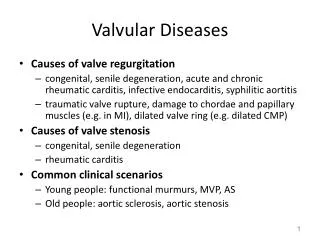
Etiology • Congenital • Acquired
Pathology • Valve Leaflet • Supporting structure
Fluid Dynamics • High-velocity jet in regurgitant orifice • Proximal acceleration • Flow disturbance in the receiving chamber • Increase volume flow across the valve
Flow Dynamics PISA High Velocity Jet Flow Disturbance
Diagnostic Approach High velocity Jet Pressure/velocity relationship of CW D
Diagnostic Approach Proximal Acceleration Proximal isovelocity Surface area
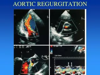
Diagnostic Approach Flow disturbance in receiving chamber Flow Mapping
Diagnostic Approach Increase volume flow across valve Volume flow at two sites.
Diagnostic Approach Increase volume flow across valve Increased antegrade velocity.
Valvular Regurgitation Volume Overload • Ventricular dilatation • Normal wall thickness
Valvular Regurgitation Total Stroke volume = blood pump by the ventricle in a single beat.
Valvular Regurgitation Forward stroke volume = the amount of blood delivered to the peripheral circulation.
Valvular Regurgitation Regurgitant volume = the amount of backflow across the abnormal valve.
Mitral Regurgitation FSV RSV
Assessment • Pulsed Doppler • CW Doppler • Color Flow Imaging • 2D imaging • M mode
Regurgitant Jet Size and Shape Factors affecting RJ Size and Shape Physiologic • Regurgitant volume. • Driving Pressure. • Shape and size of regurgitant orifice.
Regurgitant Jet Size and Shape • Receiving chamber constraint. • Wall impingement. • Timing relative to the cardiac cycle. • Influence of coexisting jets or flow-streams.
Regurgitant Jet Size and Shape Factors affecting RJ Size and Shape Technical • Ultrasound system gain • PRF • Transducer frequency
Regurgitant Jet Size and Shape • Frame rate • Image plane • Depth • Signal strength
Evaluation • Semi-quantitative • Quantitative
Semi-Quantitative Evaluation • Flow mapping (pulsed or color) • CW Doppler signal intensity • Flow reversal
Quantitative Evaluation • Volume flow at two sites • Proximal isovelocity surface area
Flow Mapping • Evaluation of the extent of the flow disturbance in the chamber receiving the regurgitant jet. • Originally performed by using pulsed Doppler. Performed now by using color Doppler.
Flow Mapping • Use multiple tomographic image plane • Note whether an abnormal flow signal with appropriate timing exist.
Flow Mapping • Integrate data from the multiple image planes. • Describe the overall 3D extent of the regurgitant jet using a scale of 0 to 4+
CW Doppler Evaluation • Compared the regurgitant signal intensity with that of the antegrade flow signal intensity. • Measure the antegrade velocity. • Evaluate the shape of the velocity curve.
Signal Intensity cm/s
Signal Intensity cm/s
Signal Intensity cm/s
Antegrade Velocity Antegrade velocity The greater the severity of the regurgitation, the higher is the antegrade velocity.
Shape of the Velocity Curve Depends on the time-varying pressure gradient across the valve.
Shape of the Velocity Curve cm/s Chronic
Shape of the Velocity Curve cm/s Acute
Upstream Reversal Severe Regurgitation at AV Valve Flow reversal in veins entering atrium
Upstream Reversal Veins Right side – Hepatic veins Left side – Pulmonary veins
Upstream Reversal D Pulmonary venous Flow S A
Upstream Reversal D Pulmonary venous Flow S Mitral Regurgitation A
Down Stream Reversal Regurgitation at the semilunar valves Flow reversal in the associated great vessels
Down Stream Reversal The distance from the valve plane that this flow reversal extends in the great vessel extends in the great vessel is proportional to regurgitant volume.
Stroke Volume Volume Flow at two Intrathoracic Sites • Total SV is calculated from antegrade flow across the regurgitant valve. • Forward SV is calculated as antegrade flow across a different valve. • Regurgitant SV = Total SV – Forward SV.
Stroke Volume SV total = CSALVOT x VTILVOT SVforward = CSALVI x VTILVI SVRegurgitant = SV total - SVforward
Stroke Volume Regurgitant Fraction RF = SVRegurgitant/ SV total
PISA • Examine the flow pattern on the upstream side of the valve. • Series of isovelocity surfaces leading to the high- velocity jet in the regurgitant orifice.