Download

1 / 49
600 likes | 1.69k Views
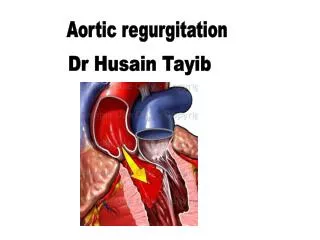
AORTIC REGURGITATION. NORMAL AORTIC VALVE. Three cusps, crescent shaped 3 commissures 3 sinuses supported by fibrous annulus 3.0 to 4.0 cm 2. Etiology of AR. LEAFLET ABNORMALITIES - Congenital bicuspid valve - Calcific valve disease -Rheumatic valve disease

E N D
NORMAL AORTIC VALVE • Three cusps, crescent shaped 3 commissures 3 sinuses supported by fibrous annulus • 3.0 to 4.0 cm2
Etiology of AR • LEAFLET ABNORMALITIES - Congenital bicuspid valve -Calcific valve disease -Rheumatic valve disease -Myxomatous valve disease -Endocarditis -Nonbacterial thrombotic endocarditis
ABNORMALITIES OF THE AORTA -Marfan syndrome -Bicuspid aortic valve disease -Hypertensive aortic dilation -Cystic medial necrosis -Aortic dissection -Systemic inflammatory diseases
FUNCTIONAL CLASSIFICATION OF AR LESIONS DYSFUNCTION ECHO FINDINGS • Enlargement of the aortic root with normal cusps • Dilatation of any components of the aortic root(aortic annulus,sinuses of valsalva,sinotubular junction)
2a:Cusp prolapse with eccentric AR jet Cusp flail Partial cusp prolapse Complete eversion of a cusp into the LVOT in long axis views Distal part of a cusp prolapsing into the LVOT(clear bending of the cusp body on long axis views and presence of a small circular structure near the cusp free edge on short axis views)
Whole cusp prolapse 2b:Free edge fenestration with eccentric AR jet Free edge of a cusp overriding the plane of aortic annulus with billowing of the entire cusp body into the LVOT(Presence of a large circular or oval structure immediately beneath the valve on short axis views) Presence of an eccentric AR jet without definite evidence of cusp prolapse
3: Poor cusp quality or quantity Thickened and rigid valves with reduced motion Tissue destruction (endocarditis) Large calcification spots/extensive calcifications of all cusps interfering with cusp motion
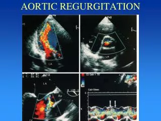
ECHO ASSESSMENT OF AR • Valve anatomy and aortic root • Assessment of LV size and function. • Doppler methods colour flow doppler Pulsed wave doppler CW Doppler
Assessment of LV size and function M-mode echo-leading edge method for linear measurements End systolic diameter End diastolic diameter 2D measurements in abnormally shaped LV. LV volume and EF-modified Simpsons method EF less reliable indicator of LV dysfunction.
COLOUR FLOW DOPPLER • REGURGITANT LESION HAS 3 COMPONENTS FLOW CONVERGANCE VENA CONTRACTA JET DIRECTION AND SIZE Jet area and jet length –not recommended Jet width Jet width/LVOT ratio>65%
Parasternal views are preffered • Jet area and length are weakly correlated with severity • Visual assessment • Over estimated in apical view • Affected by aorta to LV diastolic pressure gradient and LV compliance.
Jet width immediately below aortic valve • Proportional to the aortic valve defect • Jet width/LVOT ratio more accurate • Jet width/LVOT ratio>65% severe AR
VENA CONTRACTA • Vena contracta <3mm-mild AR, >6mm severe AR using a niquist limit of 50-60cm/sec. • Not useful in multiple jets and if the regurgitant orifice is not circular
PISA METHOD • Image the flow convergance zone in apical three or five chamber parasternal long axis upper right parasternal views Area of interest is expanded using zoom mode Color sector size is reduced to maximize the frame rate Niquist limit is adjusted to obtain a clearly visible round PISA radius
Colour flow velocity scale is shifted towards the direction jet • The PISA radius is measured as the distance between regurgitant orifice and the first aliasing in early diastole • Flow rate (Q) =2лr2хva • EROA= (6.28 X r2 X Va) / Peak V reg
PULSE WAVE DOPPLER • Holodiastolic flow reversal in decending Aorta Upper decending aorta-aortic isthmus level -suprasternal view Lower decending aorta-longitudinal subcostal view
Doppler volumetric method • Based on comparison of measurement of aortic stroke volume at the LVOT with mitral or pulmonic stroke volume. • Regurgitant volume=SV Reg valve – SV Comp Valve • RegurgitaFraction=SV Reg Valve – SV CompValve/SvReg Valve • Systemic stroke volume=LVOT area(Лd2/4=0.785d2) X LVOT VTI
AR regurgitant volume=LVOT stroke volume(total)-Mitral inflow volume(competent valve) • EROA =Regurgitant volume /TVI of regurgitant jet • Regurgitant fraction=Regurgitant volume /Total flow
CONTINUOUS WAVE DOPPLER • Signal density reflects volume of regurgitation Faint spectral display-mild AR Dense display-mod to severe AR overlap between mod to severe AR Imperfect indicator of severity
DIASTOLIC DECELERATION AND PHT • Rate of deceleration and the derived PHT reflect rate of equalizatiaon of aortic and LV diastolic pressure. • Shape of deceleration slope provides semi quantitative measure of severity of AR • Flat slope indicates mild AR • Steep slope indicates severe AR • PHT <200-severe AR,>500 –mild AR
ACUTE SEVERE AR • Infective endocraditis,aortic dissection. ECHO: Vegetations LV size normal High frequency fluttering of AML Premature closure of mitral valve diastolic MR CW doppler steep slope
Asymptomatic AR Follow up: asymptomatic mild AR,normal EF ,no LV dialatation ECHO every 2-3 yrs. In severe AR ,LV EDD 60-65mm,normal systolic function every 12 months. Every 6 months in patients with LVEDD 70mm, End systolic diameter 50mm.
MCQ 1 • According to functional classification, AR due to infective endocarditis falls into a.type1 b.type2a c.type3 d.type2b
MCQ 2 2.All are true about venacontracta in AR except a. venacontracta >6mm indicates severe AR b. useful in multiple jets c. often obtained in eccentric jets d. it provides an estimate of the size of EROA
MCQ 3 3. True about mild AR a. venacontracta <3mm b. pressure half time >500 c. EROA <10 d. regurgitant volume <40
MCQ 4 4. Severe AR is characterised by all except a. jet width/ LVOT width >65% b. regurgitant volume 60ml/beat c. regurgitant fraction >50 d. PHT <250
MCQ 5 5. True about colour flow doppler in AR are all except a. parasternal views are preferred over apical views b. jet area and length are strongly correlated with severity of AR c. jet width to LVOT ratio >65 indicates severe AR d. central jet suggestive of rheumatic disease
MCQ 6 • All are true about acute AR EXCEPT a.high velocity fluttering of AML during diastole b.LVEDP elevated c.Cwdoppler slope flat d.pulse pressure narrow
MCQ 7 • Regarding PHT all are true except a.<200 indicates severe AR b.Requires good beam alignment c.Not influenced by chamber compliance d. >500 indicate mild AR
MCQ8 • Rheumatic AR characterised by all EXCEPT a.Commissural fusion b.Commissural calcification c.Retraction of leaflets d.Produces eccentric jet
MCQ9 PISA TRUE ALL EXCEPT a.Apical view is preferred in central jet b.Apical window underestimate AR severity in eccentric jet c.EROA >30 indicates severe AR d.Regurgitant volume >50 ml indicates severe AR