Download
1 / 40
500 likes | 930 Views
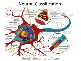
Neuron structure. fig 6-1. Myelin sheath. Peripheral nervous system: Schwann cells Central nervous system: oligodendrocytes. fig 6-2a. Myelin sheath cross section. fig 6-2c. Synapses. fig 6-5. Glial cells. fig 6-6. Glial cells. Peripheral nervous system Schwann cells: myelin sheath

E N D
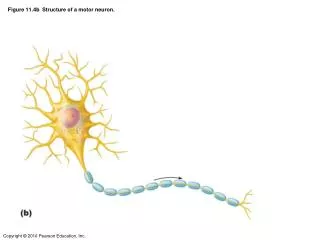
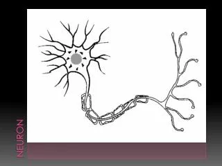
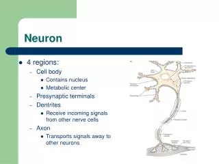
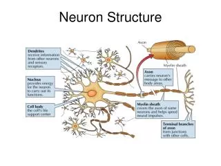
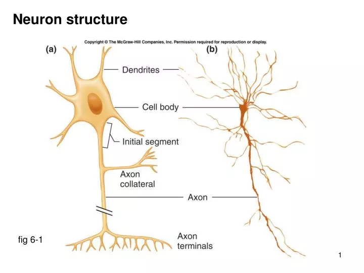
Neuron structure fig 6-1
Myelin sheath Peripheral nervous system: Schwann cells Central nervous system: oligodendrocytes fig 6-2a
Myelin sheath cross section fig 6-2c
Synapses fig 6-5
Glial cells fig 6-6
Glial cells Peripheral nervous system Schwann cells: myelin sheath Central nervous system Oligodendrocytes: myelin sheath Astrocytes: remove K+, neurotransmitters, blood-brain barrier Ependymal cells: line cavities Microglia: phagocytic Notes: 90% of CNS cells are glial cells brain tumors almost always tumors of glial cells
Classes of neurons fig 6-4 Ratios: 1 afferent : 10 efferent : 200,000 interneurons
Ion distributions; a reminder Electrochemical gradients (cell at -70 mV) Na+: strong inward (chemical: in, electrical: in) K+: weak outward (chemical: out, electrical: in) Cl-: at electrochemical equilibrium (chemical in = electrical out)
Resting membrane potential fig 6-8a Resting membrane potential of nerve cell = -70 mV
The squid axon Squid axon experiments show: Na+ & K+ gradients required anions not involved Na+/K+ pump not required if ion gradients exist energy source not required
Origin of resting membrane potential Set up: [K+]in & [Na+]out; permeability to K+ 50x permeability to Na+ Initially: K+ efflux >> Na+ influx, therefore cell negative Negative cell slows K+ efflux, speeds Na+ influx Membrane potential stabilizes when K+ efflux balances Na+ influx
Pump leak model: steady state condition Key points: only 1/10,000 ions move RMP Na+/K+ pump is electrogenic inhibition of pump (digoxin) RMP ~ -65 mV Resting Membrane Potential is predominantly a K+ diffusion potential, modulated by Na+ diffusion
Tweaking the system; altering [K+]out normal [K+]out lower [K+]out K+ gradient for efflux is steeper (more powerful) membrane potential becomes more negative i.e. “hyperpolarization” and vice versa: i.e. [K+]out depolarization
Tweaking the system; blocking Na+ channels Block Na+ influx K+ efflux continues, and RMP becomes more negative the electrical gradient for K+ influx becomes equal to the chemical gradient for K+ efflux K+ influx (electrical) = K+ efflux (chemical), i.e. equilibrium potential at which this occurs is the K+ equilibrium potential (~-90 mV)
Tweaking the system; blocking K+ channels Block K+ efflux Na+ influx continues, and potential becomes positive the electrical gradient for Na+ efflux becomes equal to the chemical gradient for Na+ influx Na+ influx (chemical) = Na+ efflux (electrical) i.e. equilibrium potential at which this occurs is the Na+ equilibrium potential (~+60 mV)
Electrochemical equilibrium; Nernst equation At equilibrium, chemical & electrical gradients are equal & opposite equation which balances chemical & electrical is Nernst pure: working: Equilibrium potentials: Na+ +60 mV K+ -89 mV Cl- -72 mV
Tweaking the system; treating with ouabain Ouabain blocks the Na+/K+ ATPase pump Na+/K+ ATPase pump moves 2K+ in and 3Na+ out; therefore the pump is “electrogenic” Ouabain treatment depolarizes cell from -70 mV to ~-65 mV Conclusion: RMP is mostly a K+ diffusion potential electrogenic Na+/K+ pump makes small direct contribution primary role of Na+/K+ pump is to set up the gradient
Polarity terminology fig 6-14
Graded potentials fig 6-15
Graded potential properties 1 can be depolarization or hyperpolarization can be large or small (i.e. graded) fig 6-16
Graded potential properties 2 shows decremental transmission Also (later, when compared with action potentials) will sum (algebraically) does not involve opening of voltage gated channels does not have threshold does not have absolute or relative refractory periods fig 6-16
Na+ channel properties fig 6-18 fig 6-20a
K+ channel properties fig 6-18 fig 6-20b
Properties of action potentials: threshold Threshold: enough voltage-gated Na+ channels open positive feedback All depend on properties of voltage-gated Na+ & K+ channels
Properties of action potentials: refractory periods Absolute refractory period: Na+ inactivation gates closed Relative refractory period: larger voltage change to reach threshold
Properties of action potentials: “all or none” When threshold is reached, complete action potential will ensue - voltage-gated Na+ & K+ channels have “programmed” sequence Action potentials; the metaphor threshold “all or none” response absolute & relative refractory period
Action potential propagation fig 6-22
Action potential propagation fig 6-22
Action potential propagation; myelin effect fig 6-23 Voltage gated Na+ & K+ channels are concentrated at the nodes of Ranvier Rates of propagation: large diameter axons faster than small diameter axons myelinated axons faster than non-myelinated axons (saltatory conduction)
Properties of graded & action potentials Property Graded potential Action potential Where PSPs, motor end plates axons, cardiac/skeletal muscle Size/polarity graded, + or - all or none Summation algebraic no (later) Threshold no yes Refractory period no absolute & relative Propagation decremental “regenerative” Properties marked depend on activity of voltage gated channels Both involve movement of very few ions i.e. (no measurable change in [ion]in & [ion]out)
Synaptic structure fig 6-26
Synaptic transmission fig 6-27
Synaptic transmission • pre-synaptic action potential (voltage-gated Na+, K+ channels) 1 • depolarization of pre-synaptic terminal 1 • opening of voltage-gated Ca++ channels 2 • Ca++ enters & binds to synaptotagmin in vesicle membrane 3 • SNARE proteins pull vesicle to docking site 4 • vesicle fusion & exocytosis of neurotransmitter (NT) 4 • neurotransmitter binds to post-synaptic receptor 5 • receptor generates post-synaptic potential • termination by re-uptake, diffusion, or degradation of NT 6 • Ca++ removed by Ca++/3Na+ counter-transport numbers 1-6 refer to Vander fig 6-27
Removal of neurotransmitters Reuptake: most important mechanism; inhibitors e.g. SSRI’s (Prozac) Diffusion away from synaptic cleft: Enzymatic destruction: only acetyl cholinesterase on AcCh inhibitors: sarin, organophosphates, physostigmine
Excitatory post synaptic potentials (EPSP’s) EPSP’s are graded potentials & have their properties EPSP’s are depolarizations of the post-synaptic membrane e.g. AcCh on skeletal muscle; opening of non-specific cation channels
Inhibitory post synaptic potentials (IPSP’s) IPSP’s are graded potentials & have their properties IPSP’s are hyperpolarizations of the post-synaptic membrane e.g. opening K+ channels; opening Cl- channels (depolarization/stabilization)
Summation of EPSP’s & IPSP’s Temporal summation: single synapse fires in rapid succession Spatial summation: separate synapses fire at ~ same time Initial segment has high concentration of voltage-gated Na+ & K+ channels site where post-synaptic potentials are converted into action potentials one EPSP produces one or more action potentials
Synaptic integration Key dendrites 2. axon 3. nucleus 4. axodendritic synapse 5. axosomatic synapse 6. myelin sheath