Download
1 / 31
380 likes | 1.39k Views
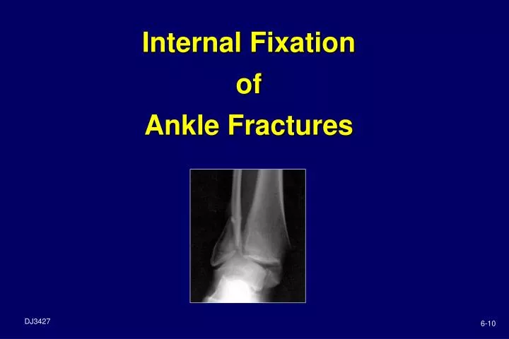
Internal Fixation of Ankle Fractures. 6-10. Objectives. Review ankle anatomy Identify the indications & treatment goals for ORIF of ankle fractures Summarize the implant options. Anatomy Ankle Bones. Formed by medial malleolus of tibia, and lateral malleolus (fibula)

E N D
Objectives • Review ankle anatomy • Identify the indications & treatment goals for ORIF of ankle fractures • Summarize the implant options
AnatomyAnkle Bones • Formed by medial malleolus of tibia, and lateral malleolus (fibula) • Talus sits in “mortise” (as in “mortise & tenon”) Fibula Tibia Talus
AnatomyAnkle Soft Tissues • Ligaments connect ankle on medial & lateral sides • Important for stability
AnatomyAnkle Soft Tissues • Fibula connected to tibia by fibrous band of tissue called syndesmosis • Also important for stability
Ankle Fractures History • Twisting injury • Immediate pain – lateral and/or medial • Difficulty weight-bearing Physical examination • Malleolar pain (posterior & anterior) • Difficulty weight-bearing • Swelling • Neurovascular involvement
Ankle FracturesRadiographs • Ankle Series: AP, mortise, lateral • “Rule out” other injuries: • Osteochondral injuries • Lateral process fracture • Anterior calcaneus fracture • Base of 5th MT fracture AP Mortise Lateral
Ankle FracturesClassification Weber / AO Classification based on level of fibula fracture A – Below syndesmosis B – At syndesmosis C – Above syndesmosis
Simple Classification: Stable & Unstable • Stable fractures • Most commonly involve medial or lateral side only • Talus remains anatomic relative to tibia
Simple Classification:Stable & Unstable • Unstable fractures • Disruption of 2 or more aspects of the mortise -- bone and/or ligament • Talus may sublux or be dislocated from tibia
Indications for SurgeryAnkle Fractures Inability to obtain or maintain an anatomic mortise (unstable fracture pattern) Open fractures
Basic Set-UpAnkle Fractures • Supine position most common • Occasionally prone for direct approach to posterior malleolus • Bump beneath ipsilateral buttocks (allows easier approach to fibula) • Tourniquet • Prep / drape to above knee • Pre-op antibiotics • Fluoroscopy or X-ray
General Considerations • Small size of ankle bones = dictates implant sizes • Multiple complex 3-D articulations • Weight bearing structure subject to high stresses (2 – 5x body weight)
General Considerations • Limited soft tissue coverage
InstrumentationAnkle Fractures • Small fragment set • Cannulated screws • K-wires • Cerclage wire • Power • Have mini-frag available
Type One malleolus Bimalleolar Tri-malleolar Treatment Fix fibula with screw / TB wire / plate Plate fibula, lag screw tibia (medial malleolus) Plate fibula, lag screw tibia, fix posterior if >20 - 25% articular surface involved Ankle FractureSurgical Tx
Implant ConsiderationsLateral Malleolus • One-third tubular plate & 3.5 mm cortex screws • Lateral • Posterior • 3.5mm compression plate for unstable fractures
Implant ConsiderationsLateral Malleolus • Locking plates -- lateral or posterolateral • Osteoporotic bone • Unstable fractures • Distal fractures
Implant ConsiderationsLateral Malleolus • Hook Plate • Used to obtain purchase in very distal fibula fractures
Posterior to anterior Anterior to posterior Implant ConsiderationsPosterior Malleolus
Implant ConsiderationsMedial Malleolus • Two partially threaded 4.0 mm cancellous screws • K-wires • Cerclage wire for tension band technique
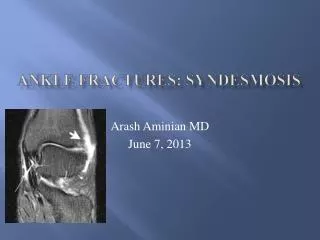
Syndesmosis FixationIndications • Syndesmotic instability after fixation of malleolus • Consider if fibula fracture > 4 cm above joint line & Maisonneuve’s fracture • Have bone hook on back table to check stability • Have large frag screws & instruments available
Implant ConsiderationsSyndesmosis • Surgeons choice of large or small fragment fully threaded screws, one or two • Not inserted as lag screw, but as a positioning screw (threads engage all cortices) • Secures position of fibula next to tibia allowing torn syndesmotic tissues to heal • May be removed in 6 - 12 weeks
Implant ConsiderationsSyndesmosis • Have pelvic forceps on back table • May need longer plates than in small frag set: • 1/3 tubular, compression or specialty fibula plate • Bioresorbable screws
Case #1 Age: 81 Gender: Female Cause of Injury: Fall Fixation: 3.5mm LCP Lateral Distal Fibula Plate
Age: 64 Gender: Female Cause of Injury: Fall Fixation: 3.5mm LCP Lateral Distal Fibula Plate Case #2
Summary • Reviewed ankle anatomy • Identified the indications & treatment goals for ORIF of ankle fractures • Summarized the implant options