Download
1 / 1
10 likes | 124 Views
Neu5Ac2-O-3Gal1-O-4GlcNAc1-O-2Man1. O. 6. Man11-O-4GlcNAc1-O-4GlcNAc-ol. Neu5Ac2-O-3Gal1-O-4GlcNAc1. 3. O. 4 2. O. Man1. O. Neu5Ac2-O-6Gal1-O-4GlcNAc1. Man a 1 – 2Man a 1. GlcNAc b 1 – 2Man a 1. 6 3. 6 4 3. Man a 1. 6 3. Man a 1. GlcNAc b 1 – .

E N D
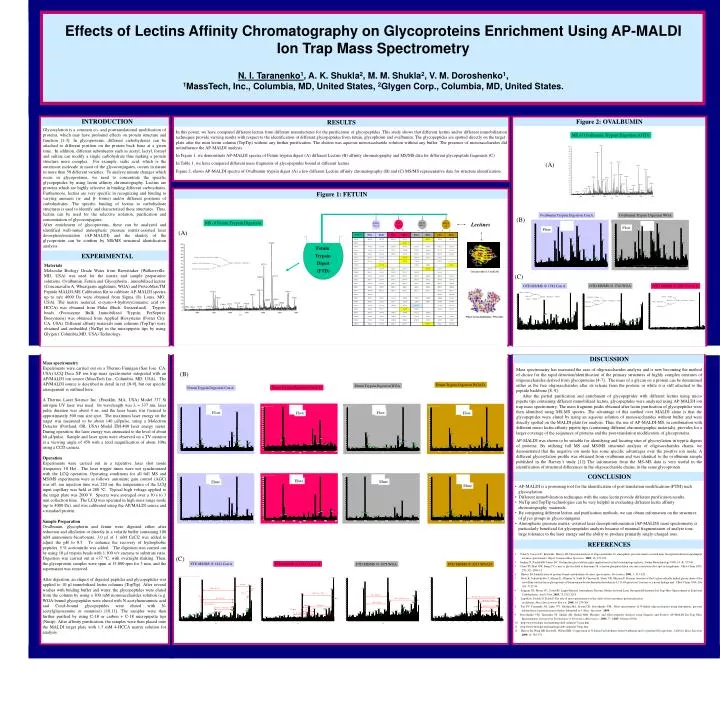
Neu5Ac2-O-3Gal1-O-4GlcNAc1-O-2Man1 O 6 Man11-O-4GlcNAc1-O-4GlcNAc-ol Neu5Ac2-O-3Gal1-O-4GlcNAc1 3 O 4 2 O Man1 O Neu5Ac2-O-6Gal1-O-4GlcNAc1 Mana1 – 2Mana1 GlcNAcb1 – 2Mana1 6 3 6 4 3 Mana1 6 3 Mana1 GlcNAcb1 – Manb1 – 4GlcNAcb1 – 4GlcNAc Manb1 – 4GlcNAcb1 – 4GlcNAc GlcNAcb1 Mana1 – 2Mana1 4 2 Mana1 GlcNAcb1 Mana1 – 3Mana1 6 4 3 GlcNAcb1 – Manb1 – 4GlcNAcb1 – 4GlcNAc Galb1 - 4GlcNAcb1 4 2 Mana1 GlcNAcb1 Effects of Lectins Affinity Chromatography on Glycoproteins Enrichment Using AP-MALDI Ion Trap Mass Spectrometry N. I. Taranenko1, A. K. Shukla2, M. M. Shukla2, V. M. Doroshenko1, 1MassTech, Inc., Columbia, MD, United States, 2Glygen Corp., Columbia, MD, United States. INTRODUCTION Figure 2: OVALBUMIN RESULTS Glycosylation is a common co- and posttranslational modification of proteins, which may have profound effects on protein structure and function [1-3]. In glycoproteins, different carbohydrates can be attached to different position on the protein back bone at a given time. In addition, different substituents such as acetyl, lactyl, formyl and sulfate can modify a single carbohydrate thus making a protein structure more complex. For example, sialic acid, which is the outermost molecule in most of the glycoconjugates, occurs in nature in more than 50 different varieties. To analyze minute changes which occur in glycoproteins, we need to concentrate the specific glycopeptides by using lectin affinity chromatography. Lectins are proteins which are highly selective in binding different carboydrates. Furthermore, lectins are very specific in recognizing and binding to varying anomers (α- and β- forms) and/or different positions of carbohydrates. The specific binding of lectins to carbohydrate structures is used to identify and characterized these structures. Thus, lectins can be used for the selective isolation, purification and concentration of glycoconjugates. After enrichment of glycoproteins, these can be analyzed and identified well-suited atmospheric pressure matrix-assisted laser desorption/ionization (AP-MALDI) and the identity of the glycoprotein can be confirm by MS/MS structural identification analysis. In this poster, we have compared different lectins from different manufactures for the purification of glycopeptides. This study shows that different lectins and/or different immobilization techniques provide varying results with respect to the identification of different glycopeptides from fetuin, glycophorin and ovalbumin. The glycopeptides are spotted directly on the target plate after the mini lectin column (TopTip) without any further purification. The elution was aqueous monosaccharide solution without any buffer. The presence of monosaccharides did notinfluence the AP-MALDI analysis. In Figure 1, we demonstrate AP-MALDI spectra of Fetuin trypsin digest (A) different Lectins (B) affinity chromatography and MS/MS data for different glycopeptide fragments (C) . In Table 1, we have compared different mass fragments of glycopeptides bound at different lectins. Figure 2, shows AP-MALDI spectra of Ovalbumin trypsin digest (A) a few different Lectins affinity chromatography (B) and (C) MS/MS representative data for structure identification. MS of Ovalbumin Trypsin Digestion (OTD) (A) Figure 1: FETUIN Ovalbumin Trypsin Digestion Con A Ovalbumin Trypsin Digestion WGA (B) MS of Fetuin Trypsin Digestion Lectines Sigma WGA Sigma Con A Con A-D WGA D Flow Flow (A) Fetuin Trypsin Digest (FTD) EXPERIMENTAL Materials Molecular Biology Grade Water from Biowittaker (Walkersville, MD, USA) was used for the matrix and sample preparation solutions. Ovalbumin, Fetuin and Glycophorin , immobilized lectins (Concanavalin A, Wheatgerm agglutinin, WGA) and ProteoMassTM Peptide MALDI-MS Calibration Kit to calibrate AP-MALDI spectra up to m/z 4000 Da were obtained from Sigma (St. Louis, MO, USA). The matrix material, α-cyano-4-hydroxycinnamic acid (4-HCCA) was obtained from Fluka (Buch, Switzerland). Trypsin beads (Poroszyme Bulk Immobilized Trypsin, PerSeptive Biosystems) was obtained from Applied Biosystems (Foster City, CA, USA). Different affinity materials mini columns (TopTip) were obtained and embedded (NuTip) in the micropipette tips by using Glygen ( Columbia,MD, USA) Technology. Concanavalin A: ConA [10] (C) OTD MS/MS @ 1746 WGA OTD MS/MS @ 1582 Con A OTD MS/MS @ 1867 Con A-D Wheat Germ Agglutinine: WGA [11] DISCUSSION Mass spectrometry Experiments were carried out on a Thermo Finnigan (San Jose, CA, USA) LCQ Deca XP ion trap mass spectrometer integrated with an AP/MALDI ion source (MassTech Inc., Columbia, MD, USA). The AP/MALDI source is described in detail in ref. [8-9], but our specific arrangement is outlined here. A Thermo Laser Science Inc. (Franklin, MA, USA) Model 337 Si nitrogen UV laser was used. Its wavelength was l = 337 nm, laser pulse duration was about 4 ns, and the laser beam was focused to approximately 500 mm size spot. The maximum laser energy on the target was measured to be about 140 mJ/pulse, using a Molectron Detector (Portland, OR, USA) Model EM-400 laser energy meter. During operation, the laser energy was attenuated to the level of about 60 mJ/pulse. Sample and laser spots were observed on a TV monitor at a viewing angle of 450 with a total magnification of about 100x using a CCD camera. Operation Experiments were carried out in a repetitive laser shot mode (frequency 10 Hz). The laser trigger times were not synchronized with the LCQ operation. Operating conditions for all full MS and MS/MS experiments were as follows: automatic gain control (AGC) was off, ion injection time was 220 ms, the temperature of the LCQ input capillary was held at 280 0C. Typical high voltage applied to the target plate was 2000 V. Spectra were averaged over a 30 s to 3 min collection time. The LCQ was operated in high mass range mode (up to 4000 Da), and was calibrated using the AP/MALDI source and a standard protein. Sample Preparation Ovalbumin, glycophorin and fetuin were digested either after reduction and alkylation or directly in a volatile buffer containing 100 mM ammonium bicarbonate. 3.0 µl of 1 mM CaCl2 was added to adjust the pH to 8.5. To enhance the recovery of hydrophobic peptides, 5 % acetonitrile was added. The digestion was carried out by using 10 µl trypsin beads with 1:100 v/v enzyme to substrate ratio. Digestion was carried out at +37 0C, with overnight shaking. Then, the glycoprotein samples were spun at 15,000 rpm for 3 min. and the supernatant was removed. After digestion, an aliquot of digested peptides and glycopeptides was applied to 10 µl immobilized lectin columns (TopTip). After several washes with binding buffer and water, the glycopeptides were eluted from the column by using a 300 mM monosaccharides solution (e.g. WGA-bound glycopeptides were eluted with N-acetylneuraminic acid and ConA-bound glycopeptides were eluted with N-acetylglucosamine or mannose) [10,11]. The samples were then further purified by using C-18 or carbon + C-18 micropipette tips (Nutip). After affinity purification, the samples were then placed onto the MALDI target plate with 1.5 mM 4-HCCA matrix solution for analysis. Mass spectrometry has increased the ease of oligosaccharides analysis and is now becoming the method of choice for the rapid detection/identification of the primary structures of highly complex mixtures of oligosaccharides derived from glycoproteins [4-7]. The mass of a glycan on a protein can be determined either as the free oligosaccharides after its release from the protein, or while it is still attached to the peptide backbone [8, 9]. After the partial purification and enrichment of glycopeptides with different lectins using micro pipette tips containing different immobilized lectins, glycopeptides were analyzed using AP-MALDI ion trap mass spectrometry. The mass fragment peaks obtained after lectin purification of glycopeptides were then identified using MS-MS spectra. The advantage of this method over MALDI alone is that the glycopeptides were eluted by using an aqueous solution of monosaccharides without buffer and were directly spotted on the MALDI plate for analysis. Thus, the use of AP-MALDI-MS, in combination with different micro lectin affinity pipette tips (containing different chromatographic materials), provides for a larger coverage of the sequences of proteins and the post-translation modification of glycoproteins. AP-MALDI was shown to be suitable for identifying and locating sites of glycosylation in tryptic digests of proteins. By utilizing full MS and MS/MS structural analysis of oligosaccharides chains, we demonstrated that the negative ion mode has some specific advantages over the positive ion mode. A different glycosylation profile was obtained from ovalbumin and was identical to the ovalbumin sample published in the Harvey’s study [12] The information from the MS-MS data is very useful in the identification of structural differences in the oligosaccharide chains, in the same glycoprotein. (B) Fetuin Trypsin Digestion WGA D Fetuin Trypsin Digestion WGA Fetuin Trypsin Digestion Con A-D Fetuin Trypsin Digestion Con A Flow Flow Flow Flow CONCLUSION Elute Elute Elute Elute • AP-MALDI is a promising tool for the identification of post translation modifications (PTM) such • glycosylation. • Different immobilization techniques with the same lectin provide different purification results. • NuTip and TopTip technologies can be very helpful in evaluating different lectin affinity • chromatography materials. • By comparing different lectins and purification methods, we can obtain information on the structures • of glyco groups in glycoconjugates. • Atmospheric pressure matrix- assisted laser desorption/ionization (AP-MALDI) mass spectrometry is • particularly beneficial for glycopeptides analysis because of minimal fragmentation of analyte ions, • large tolerance to the laser energy and the ability to produce primarily singly-charged ions. REFERENCES 1.Colin S, Creaser JC, Reynolds, Harvey DJ. Structural analysis of oligosaccharides by atmospheric pressure matrix-assisted laser desorption/ionization quadrupole ion mass spectrometry. Rapid Commun.Mass Spectrom. 2002; 16, 176-184. 2. Jenkins N, Parekh RB, James DC. Getting the glycosylation right: implications for the biotechnology industry. Nature Biotechnology 1996; 14 (8): 975-81. 3. Chou TY, Hart GW, Dang CV. c-myc is glycosylated at threonine-58, a known phosphorylation site and a mutational hot-spot in lymphomas. J Biol Chem 1995; 270 (32): 18961-5. 4. Harvey DJ, Identification of protein-bound carbohydrates by mass spectrometry. Proteomics.2001, 1, 311-328. 5. Bock K, Schusterkolbe J, Altman E, Allmaier G, Stahl B, Christian R, Sleytr UB, Messner P. Primary structure of the O-glycosidically linked glycan chain of the crystalline surface-layer glycoprotein of thermoanaerobacter-thermohydrosufuricus L111-69-galactosyl tyrosine as a novel linkage unit. J Biol Chem 1994; 269 (10): 7137-44. 6. Seggern CE, Moyer SC, Cotter RJ. Liquid Infrared Atmospheric Pressure Matrix-Assisted Laser Desorption/Ionization Ion Trap Mass Spectrometer of Sialylated Carbohydrates. Anal Chem.,2003, 75,3212-3218. 7. Lapolla A, Fedele D, Traldi P. The role of mass spectrometry in the study of non-enzymatic protein glycation in diabetes. Mass Spectrometry Rewiew,2000, 19, 279-304. 8. Tan PV, Taranenko NI, Laiko VV, Yakshin MA, Prasad CR, Doroshenko VM. Mass spectrometry of N-linked oligosaccharides using atmospheric pressure infrared laser ionization from solution. Submitted to J. Mass Spectrom. 2004. 9. Doroshenko VM, Taranenko NI, Shukla AK, Shukla MM. Phospho- and Glyco-peptides Analysis using Negative and Positive AP-MALDI Ion Trap Mass Spectrometery. Intergrating Technologies in Proteomics &Genomics. 2004, 77, ABRF, Oregon (2004). 10. http://www.biologie.uni-hamburge.de/b-online/e17/cona.htm 11. http://www.biologie.uni-hamburge.de/b-online/e17/wga.htm 12. Harvey Dj, Wing DR, Kuster B, Wilson IBH. Composition of N-Liked Carbohydrates from Ovalbumin and Co-purified Glycoproteins. J.AM Soc Mass Spectrom2000, 11, 564-571. (C) FTD MS/MS @ 1621 Con A FTD MS/MS @ 1783 Con A-D FTD MS/MS @ 1978 WGA FTD MS/MS @ 3273 WGA D (NeuSGc) (Hex) (NeuAc) -H2O (HexNAc)2 + TFA CH3 - C (Hex)(NeuAc) -H2O (HexNAc) (Hex)2 (NeuAc)2 less H2O (Hex) (NeuAc) (Hex)2 (NeuAc less Ac) -H2O (HexNAc)2 (NeuAc) (Hex)(NeuAc) (Hex) (NeuGc) (HexNAc)2 TFA (Hex) (HexNAc) (NeuAc) less Co 2370.2 (NeuAc) or (NeuGc) – (H2O) 1127.6 (HexNAc) (Hex)3 (NeuAc) (HexNAc) (Hex) (NeuAc)