Biological Membranes
340 likes | 738 Views
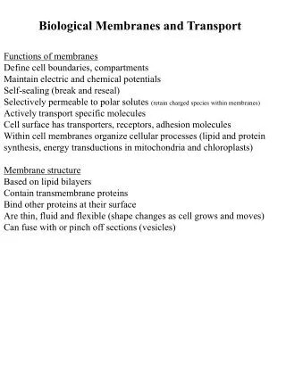
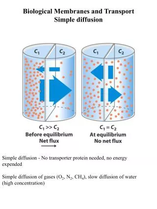
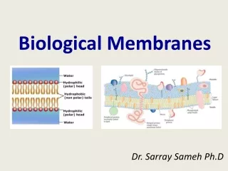
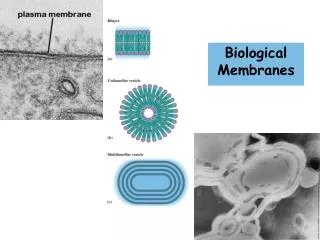
Biological Membranes. Amphipathic lipid aggregates that form in water . Monolayer of oil molecules at an air-water interface. Bilayers are noncovalent, cooperative structures. Membrane Phase Transitions. The "melting" of membrane lipids

Biological Membranes
E N D
Presentation Transcript
Amphipathic lipid aggregates that form in water Monolayer of oil molecules at an air-water interface Bilayers are noncovalent, cooperative structures
Membrane Phase Transitions The "melting" of membrane lipids • Below a certain transition temperature, membrane lipids are rigid and tightly packed • Above the transition temperature, lipids are more flexible and mobile • The transition temperature is characteristic of the lipids in the membrane From Lehninger Principles of Biochemistry
Higher the proportion of saturated fatty acid, higher is the transition temperature. From Lehninger Principles of Biochemistry
Sterol content of a membrane has 2 effects on membrane fluidity Below the transition temperature: Insertion of rigid planar sterol prevents highly ordered packing of fatty acid side chains Membrane fluidity Above the transition temperature: Rigid planar sterol reduces the freedom of neighboring fatty acid side chains Membrane fluidity
Cells regulate their lipid composition to achieve a constant membrane fluidity under various growth conditions From Lehninger Principles of Biochemistry
Motion of Membrane Lipids Lateral Diffusion Transbilayer or flip-flop Diffusion From Lehninger Principles of Biochemistry
Flippases A relatively new discovery! • Lipids can be moved from one monolayer to the other by flippase proteins • Some flippases operate passively and do not require an energy source • Other flippases appear to operate actively and require the energy of hydrolysis of ATP From Garrett & Grisham
Singer & Nicolson defined two classes • Integral (intrinsic) proteins • Peripheral (extrinsic) proteins • We'll note a new one – lipid-anchored proteins
A. Peripheral, Integral & Lipid-Linked Proteins C. B. From Lehninger Principles of Biochemistry
Lipid-linked membrane proteins Covalently attached lipids anchor membrane proteins to the lipid bilayer A relative new class of membrane proteins 4 types have been found: Amide-linked myristoyl anchors Thioester-linked fatty acyl anchors Thioether-linked prenyl anchors Glycosyl phosphatidylinositol anchors Glycosyl phosphatidylinositol (GPI) anchor From Lehninger Principles of Biochemistry
Some membrane proteins span the lipid bilayer Glycophorin in the erythrocyte A single-transmembrane-segment protein • One transmembrane segment with globular domains on either end • Transmembrane segment is alpha helical and consists of 19 hydrophobic amino acids • Extracellular portion contains oligosaccharides (and these constitute the ABO and MN blood group determinants)
Hydropathy Plots 1 From Lehninger Principles of Biochemistry
Demonstration of lateral diffusion of membrane proteins Membrane proteins, like membrane lipids, are free to diffuse laterally in the plane of the bilayer
Integral Membrane Proteins Held in the membrane by hydrophobic interactions with lipids From Lehninger Principles of Biochemistry
Asymmetric distribution of phospholipids between the inner & outer monolayers of erythrocyte plasma membrane
Bacteriorhodopsin, a membrane-spanning protein Bound retinal From Lehninger Principles of Biochemistry
3-D structure of the photosynthetic reaction center of purple bacterium First integral membrane protein to have its structure determined by X-ray diffraction methods Prosthetic group (light-absorbing pigments) Residues that are part of the trans-membrane helices From Lehninger Principles of Biochemistry
Porin FhuA, an integral membrane protein with b-barrel structure Not all integral membrane proteins are composed of transmembrane a helices Porin allows certain polar solutes to cross the outer membrane of bacteria From Lehninger Principles of Biochemistry
Porins Found both in Gram-negative bacteria and in mitochondrial outer membrane • Porins are pore-forming proteins (30-50 kD) • Most arrange in membrane as trimers • High homology between various porins • Porin from Rhodobacter capsulatus has 16-stranded beta barrel that traverses the membrane to form the pore
Porins are transmembrane channels for small molecules 22-stranded b barrel (seen as a hollow pipe) Cork domain (keeps the channel closed) Binding of ferrichrome-iron on the outer surface causes conformation change & moves the ferrichrome into the barrel Structure of FhuA, an iron transporter from E.coli From Lehninger Principles of Biochemistry
Gap Junctions Vital connections for animal cells • Provide metabolic connections • Provide a means of chemical transfer • Provide a means of communication • Permit large number of cells to act in synchrony (for example, synchronized contraction of heart muscle is brought about by flow of ions through gap junctions)
Induces closure of gap junction central channel Gap Junctions • Hexameric arrays of a single 32 kD protein • Subunits are tilted with respect to central axis • Pore in center can be opened or closed by the tilting of the subunits, as response to stress
Aquaporins form hydrophilic transmembrane channels for the passage of water Proposed structure of aquaporin channel (Formed by 4 monomers) Likely transmembrane topology of an aquaporin, AQP-1 Monomer Water flows through the channel in single file at the rate of 5 X 10⁸ molecules / second From Lehninger Principles of Biochemistry