Download

1 / 18
310 likes | 1.56k Views
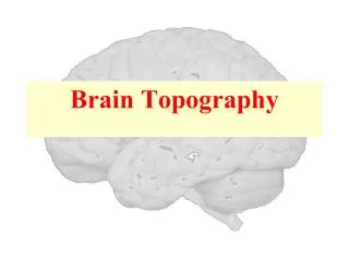
Brain Topography. Dr. Nimir Dr. Safaa. Objectives Demarcate the major lobes, gyri and sulci of the cerebral hemisphere . Describe the organization of the cerebral hemisphere into cerebral cortex ,white matter and nuclei

E N D
Brain Topography • Dr. Nimir • Dr. Safaa
Objectives • Demarcate the major lobes, gyri and sulci of the cerebral hemisphere. • Describe the organization of the cerebral hemisphere into cerebral cortex ,white matter and nuclei • Describe the types of fibers in the white matter of the cerebral hemisphere: projection (internal capsule), commissural and association fibers.
The brain is divided into: • 1- Forebrain (Telencephalon= cerebrum & diencephalon). • 2- Midbrain. • 3- Hindbrain (pons ,medulla oblongata, cerebellum). • Cerebrum (cerebral hemispheres & basal ganglia ). • Cerebral hemispheres are seperated by longitudinal fissure into left & right which are connected by corpus callosum.
Each cerebral hemisphere has 3 surfaces: • Superolateral. • Medial. • Inferior. • Each cerebral hemisphere has 3 poles: • Frontal pole. • Temporal pole. • Occipital pole.
To increase the surface area the cerebral cortex is thrown into folds or gyri, which are separated from each other by sulci or fissures. • Each cerebral hemisphere is divided by central, parieto-occipital,lateral and calcarine sulci into frontal, parietal, temporal, and occipital lobes.
Superolateralsurface • Frontal lobe: • Is anterior to the central sulcus and superior to the lateral sulcus. • Its superolateral surface is divided by three sulciinto four gyri. • Precentralsulcusruns parallel to central sulcus, and precentralgyruslies between them. • Extending anteriorly from precentralsulcus are superior and inferior frontal sulci. • Superior frontal gyruslies above superior sulcus. • Middle frontal gyruslies between superior and inferior sulci. • Inferior frontal gyruslies below inferior sulcus.
Parietal lobe: • Is posterior to central sulcus, superior to lateral sulcusand anterior to parieto-occipital sulcus. • Its lateral surface is divided by two sulci into three gyri. • Postcentralsulcus runs parallel to central sulcus, and postcentralgyruslies between them. • Running posteriorly from the middle of the postcentral sulcus is intraparietalsulcus. Superior to intraparietal sulcus is superior parietal lobule (gyrus). • Inferior to the intraparietal sulcus is the inferior parietal lobule (gyrus).
Temporal lobe: • Is inferior to the lateral sulcus. • Its lateral surface is divided into three gyri by two sulci. • The superior and middle temporal sulci run parallel to lateral sulcus and divide temporal lobe into superior, middle, and inferior temporal gyri. • Occipital lobe: • Is behind the parieto-occipital sulcus.
Medial surfaces • Corpus callosum is the largest commissure. • Cingulate gyrusis above corpus callosum,separatedfrom it by callosalsulcus and from medial frontal gyrusby cingulate sulcus. • Paracentral lobule anteriorly is part of precentralgyrusposteriorly is part of postcentralgyrus. • Precuneus is part of parietal lobe. • Cuneuswhich is part of occipital lobe, is between parieto-occipita & calcrinesulci.
Inferior surface • Collateral sulcus runs anteriorly below the calcarine sulcus. • Between collateral sulcus and calcarine sulcus is lingual gyrus. • Anterior to lingual gyrus is parahippocampalgyrus which terminates in front as uncus. • Medial occipitotemporalgyrusis bounded medially by collateraland rhinal sulci and laterally by occipitotemporalsulcus which separate it from lateral occipitotemporalgyrus. • On inferior surface of frontal lobe is olfactory sulcuswith olfactory bulb and orbital gyri.
Gray matter consists of nerve cells(neurons) embedded in neuroglia. • White matter consists of nerve fibers (axons) embedded in neuroglia. • In cerebral hemispheres gray mater is seen in cerebral cortex & basal ganglia.
White mater fibers are classified into three groups according to their connections: • 1. Commissural fibers. • 2. Association fibers. • 3. Projection fibers.
1. Commissural fibers: • Connect corresponding regions of the two hemispheres. They are: • Corpus callosum(splenium ,body, genu, rostrum). • Anterior commissure. • Posterior commissure. • Fornix. • Habenularcommissure.
2. Association fibers: • Connect various cortical regions within the same hemisphere and may be divided into short and long. • Shortassociation fibers connect adjacent gyri. • Longassociation fibers: • Uncinatefasciculus connects motor speech area & inferior surface of the frontal lobe to temporal lobe.
Cingulumwithin cingulate gyrus connects frontal and parietal lobes to parahippocampal and temporal cortical regions. • Superior longitudinal fasciculus connects anterior part of the frontal lobe to the occipital and temporal lobes. • Inferior longitudinal fasciculus connects occipital lobe to temporal lobe. • Fronto-occipital fasciculus connects frontal lobe to occipital and temporal lobes.
3. Projection fibersare afferent and efferent nerve fibers passing to and from the brainstem & spinal cord to the entire cerebral cortex. • Mainly pass within the internal capsule between caudate nucleus &thalamus medially &lentiformnucleus laterally. • The fibers radiate to cortical areas as corona radiata.